Protecting damaged hearts with microRNAs

Once the heart is fully formed, the cells that make up heart muscle, known as cardiomyocytes, have very limited ability to reproduce themselves. After a heart attack, cardiomyocytes die off; unable to make new ones, the heart instead forms scar tissue. Over time, this can set people up for heart failure.
New work published April 17th in Nature Communications advances the possibility of reviving the heart's regenerative capacities using microRNAs—small molecules that regulate gene function and are abundant in developing hearts.
In 2013, Da-Zhi Wang, Ph.D., a cardiology researcher at Boston Children's Hospital and a professor of pediatrics of Harvard Medical School, identified a family of microRNAs called miR-17-92 that regulates proliferation of cardiomyocytes. In new work, his team shows two family members, miR-19a and miR-19b, to be particularly potent and potentially good candidates for treating heart attack.
Short- and long-term protection
Wang and colleagues tested the microRNAs delivered two different ways. One method gave them to mice directly, coated with lipids to help them slip inside cells. The other method put the microRNAs into a gene therapy vector designed to target the heart.
Injected into mice after a heart attack—either directly into the heart or systemically—miR-19a/b provided both immediate and long-term protection. In the early phase, the first 10 days after heart attack, the microRNAs reduced the acute cell death and suppressed the inflammatory immune response that exacerbates cardiac damage. Tests showed that these microRNAs inhibited multiple genes involved in these processes.
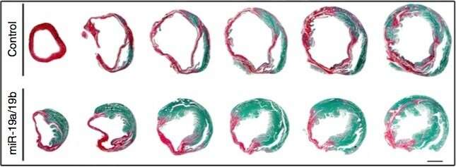
Longer-term, the treated hearts had more healthy tissue, less dead or scarred tissue and improved contractility, as evidenced by increased left-ventricular fractional shortening on echocardiography. Dilated cardiomyopathy—a stretching and thinning of the heart muscle that ultimately weakens the heart—was also reduced.
"The initial purpose is to rescue and protect the heart from long-term damage," says Wang. "In the second phase, we believe the microRNAs help with cardiomyocyte proliferation."
One and done?
Aside from regulating multiple genetic targets, microRNAs have another advantage as a therapy: unlike gene therapy, they don't linger in the heart.
"They go in very fast and do not last long, but they have a lasting effect in repairing damaged hearts," says Jinghai Chen, Ph.D., a former member of the Wang lab and co-corresponding author on the paper with Wang. (Chen is now on the faculty at the Institute of Translational Medicine, Zhejiang University, Hangzhou, China.) "We gave mice only one shot when the heart needed the most help, then so we kept checking expression level of miRNA19a/b post-injection. After one week, expression decreased to a normal level, but the protection lasted for more than one year."
Even when given systemically, the microRNAs tended to go to the site of heart damage. But Wang would like to optimize the specificity of the treatment, since the miRNAs can also affect other tissue and organs. The next step would be to test that treatment in a larger animal before advancing to studies in humans.
All of us make miR-19a/b to some degree, so the treatment would be boosting something we already have. "MicroRNAs hold tremendous promise to become powerful tools to battle cardiovascular disease," the researchers write.
More information: Feng Gao et al, Therapeutic role of miR-19a/19b in cardiac regeneration and protection from myocardial infarction, Nature Communications (2019). DOI: 10.1038/s41467-019-09530-1


